|
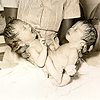
Identical twins develop when a single fertilized egg, also known as a monozygote, splits during the first two weeks of conception. Conjoined twins form when this split occurs after the first two weeks of conception. The monozygote does not fully separate and eventually develops into a conjoined fetus that shares one placenta, one amniotic sac, and one chorionic sac. Because the twins develop from a single egg, they will also be the same sex. The extent of separation and the stage at which it occurs determine the type of conjoined twin, i.e., where and how the twins will be joined.
The diagram at left illustrates the germ layers in embryos of conjoined twins. The proximity of the segments determines how much shared tissue there will be. The further apart the segments, the greater the likelihood that the organs will develop fully in each fetus. If the segments are at their farthest point, there will only be a minimum of tissue and cartilage joining the twins, i.e. omphalopagus twins will develop. Classification of Conjoined TwinsIsidore Geoffroy Saint-Hillaire was the first teratologist to classify conjoined twins, using Greek etymology to describe the twins in terms of their shared anatomy. Many of his terms are still in use today. Listed below are some commonly occurring types of conjoined twins, examples of which may be viewed throughout this exhibit.
Craniopagus: joined at the cranium (head).
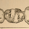
Omphalopagus or xiphopagus: joined in the region of the umbilicus.
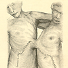
Thoracopagus: joined at the thoracic cavity (chest).
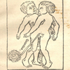
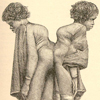
Ischiopagus: joined at the inferior margins of the coccyx and sacrum, with two separate spinal columns.
Pygopagus: joined at the lateral and posterior surfaces of the coccyx and sacrum. (责任编辑:) |